
The Sacrum. Our body’s sacred foundation
The Sacred Bone, Trauma & Nervous System Support.
CRANIOSACRAL THERAPY & SCAR WORK IN NERVOUS SYSTEM REGULATION.

The sacrum is a triangular bone at the base of the spine, resting between the hips, and yet its role reaches far beyond anatomy. Often described as the sacred bone, the sacrum forms the foundation of our posture, movement, and sense of support. In craniosacral and somatic therapy, the sacrum is understood not only as a structural centre, but as a place where the nervous system listens for safety. When the sacrum feels held, the body often responds with ease, grounding, and a subtle sense of being supported from within.
Connecting with your sacrum, a gentle somatic exercise
You can try this seated or lying down.
Begin by noticing where your body meets the surface beneath you.
Without changing anything, bring gentle awareness to the area at the base of your spine, the place where your weight is naturally received.
You might place one hand over your lower back or pelvis, simply as an invitation.
Take a slow breath in, and allow the exhale to soften downward, as if the sacrum could rest a little more heavily.
There is nothing to achieve here. No correction. Just sensing.
Notice if the body responds, perhaps with warmth, a deeper breath, or a feeling of settling.
If nothing happens, that is also welcome.
This quiet contact with the sacrum can be a first step toward grounding, regulation, and a felt sense of support, a gentle reminder that the body already knows how to be held.
The Name
If the historical sources are to be believed, the English term sacrum entered the anatomical lexicon in the mid-eighteenth century, derived from the Latin os sacrum — meaning the holy bone.
Why holy?
Some traditions suggest that, as a centre of reproductive power, the sacrum earned its sacred status through its association with creation, grounding, and what many would recognise as the root chakra. Others point to ancient records describing the sacral bone’s role in ritual practices, their deeper meaning now largely lost to time.
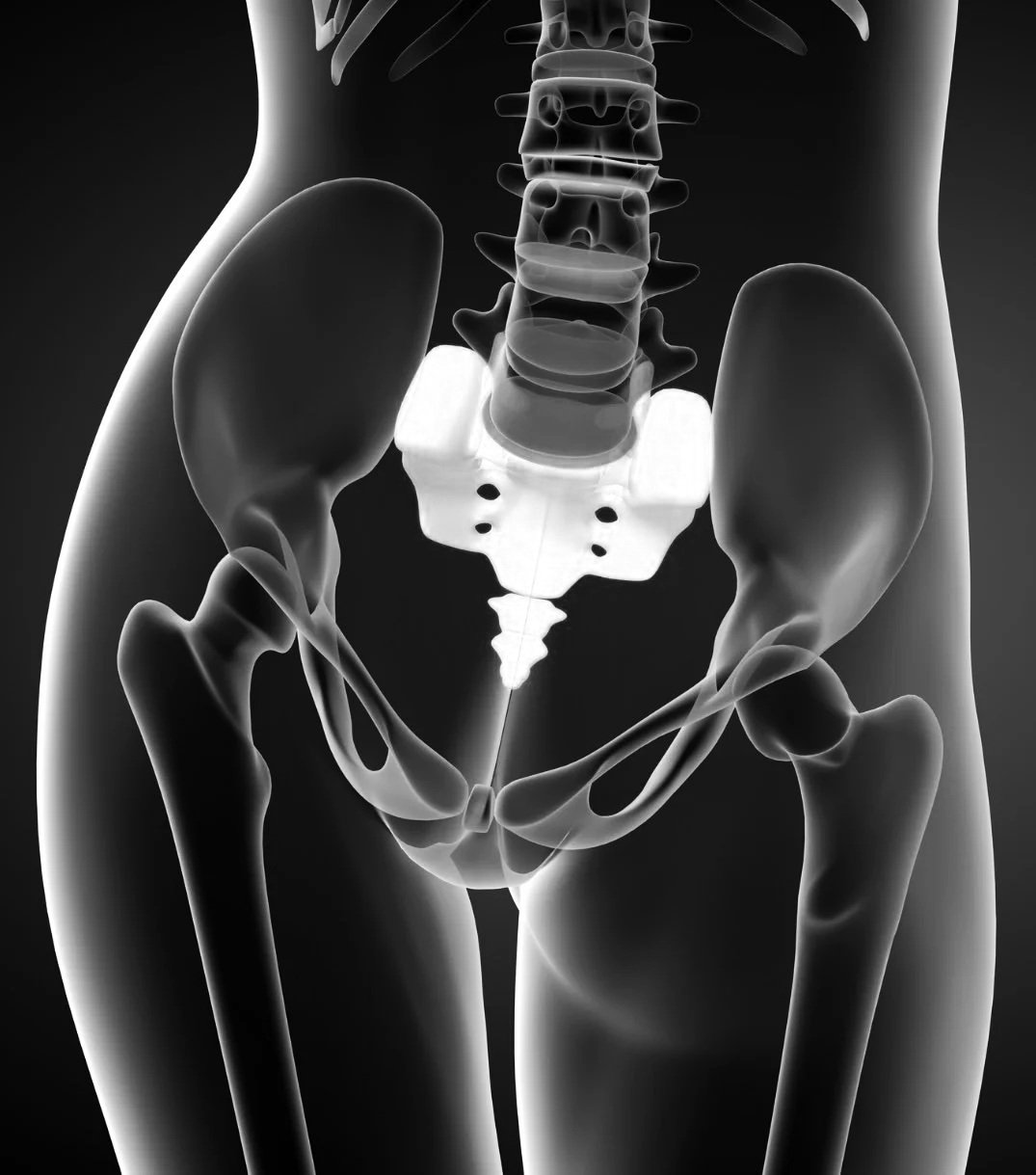
Articulations
The sacrum is wide at the top and narrow at the bottom. This triangular shape is ‘wedged' between:
The lowest part of the lumbar spine - L5 vertebrae
The two ilium bones, i.e., the large bones at the side of your hips; and
The coccyx, also known as tailbone, located at the very end of the spine (more on that truly under-appreciated bone and its importance later).
Located in between such strong neighbours, sacrum is sensitive to weight. How?
The more weight we carry, and I am not talking about grocery shopping here, but the extra ‘cushioning' that we accumulate in our bodies over the years, the harder the sacrum’s life becomes. The extra weight puts an additional downward pressure on it. The months or years of such extra pressure might translate into tension between the L5, the hips and the coccyx.
Structure
The sacrum is composed of five segments (also known as S1-S5). They start their life as properly separated vertebrae, and then gradually fuse together over time. The fusion process starts as early as eight weeks following conception, and continues until the age of 25. Some say that the fusion speeds up greatly from the age of 18 onwards. Once we reach 25, our sacrum is fused and we are done growing (hopefully only in a physical sense though).
However, the intervertebral discs that were originally between the sacral segments are still there, and they stay there well into our elderly years. We call these formations intervertebral disk material. Each 1.5 mm layer is flattened, but still present, cushioning and shock-absorbing, and thus making the sacrum the strongest bone in a body.
Fun fact - the level of sacral segment fusion is one of the factors used in forensic investigation in order to establish the age of a body found in suspect circumstances.
Important Connections:
Symphysis Pubis
The pubic symphysis is the cartilage connecting the two sides of the pelvis. Due to hormonal changes in pregnancy, it softens allowing the pelvis to open during birth. It is an important factor in sacral health and movement, despite not being “directly” connected to the sacrum.
The symphysis pubis gradually grows harder with age. With passing time, its composition changes from cartilage (soft, bouncy, pliable) to bone (hard and allowing very little movement). It influences the sacrum greatly because it inhibits its movement. We shouldn’t despair though. On average, we have until our 70s before this change is fully in place. Interestingly, keeping the pelvis flexible can buy us a few extra years of freedom of movement. And if you are flexible enough to do splits at the ripe age of 70, your symphysis pubis may never harden.
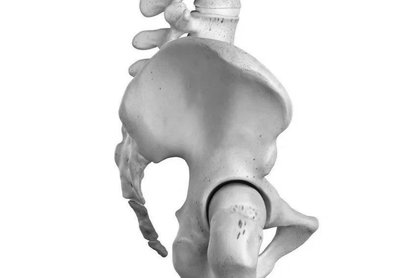
2. Sacroiliac Joints
The sacroiliac joints are L-shaped structures that play a crucial role in stability, movement, and load transfer between the spine and the pelvis. Though small in range, their influence is significant.
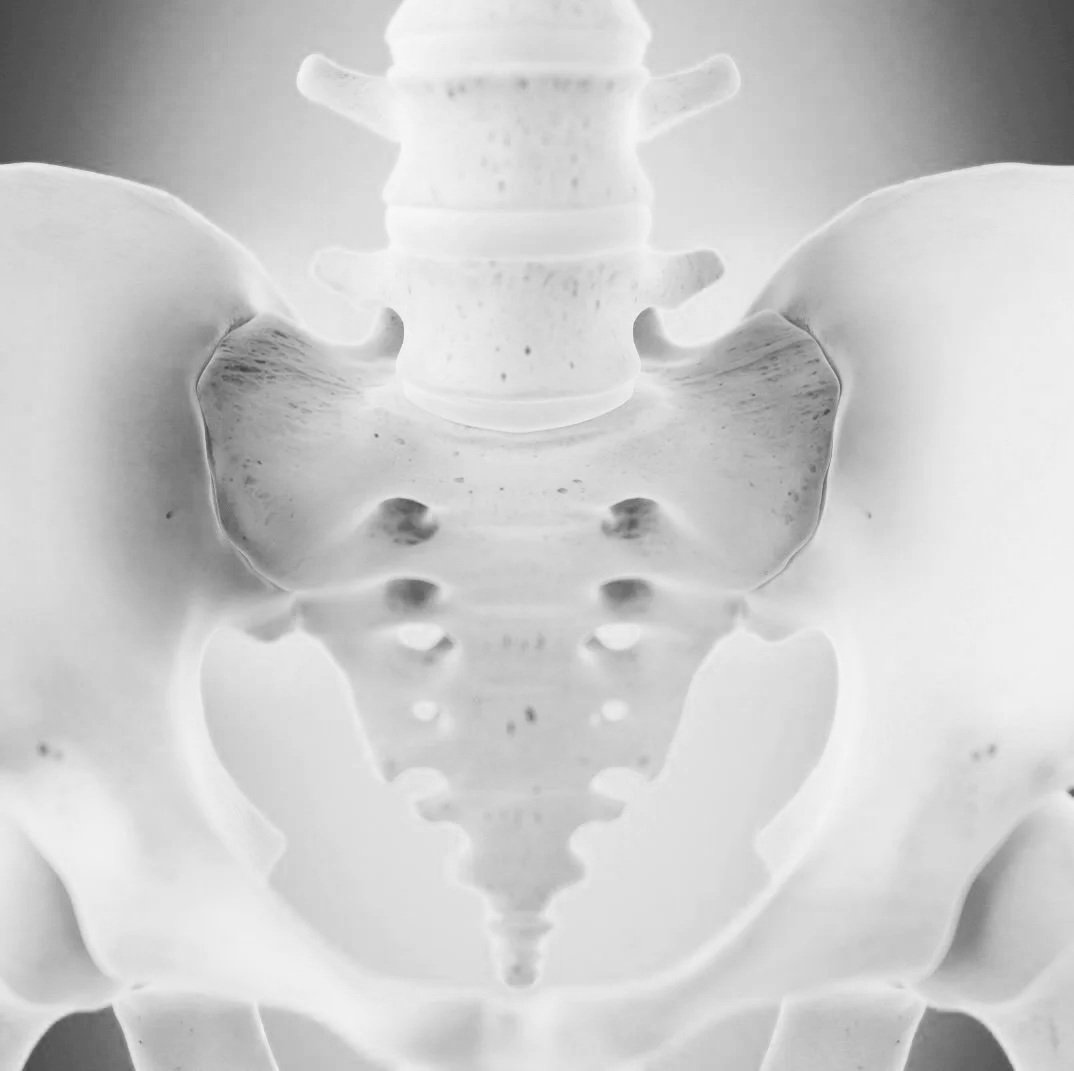
You may recognise the subtle dimples visible in the lower back of some people, these mark the level of the first sacral segment, close to where the sacroiliac joints sit. Often, the sacroiliac joints only come into awareness when discomfort arises, particularly in cases of lower back or pelvic pain. When tension, imbalance, or restriction is present in this area, it can affect both movement and overall ease in the body.
As is often the case in anatomy, the name describes the relationship. The sacroiliac joints connect the first and second sacral segments, and part of the third, to the iliac bones of the pelvis, sometimes poetically referred to as the wings of the pelvis. Through this connection, the sacrum becomes a central bridge between the upper and lower body, supporting both structural integrity and fluid motion.
3. The Coccyx
The coccyx is formed by four fused vertebrae and completes the length of the spine. Extending from the base of the sacrum, it sits in close proximity to the pelvic floor and urogenital structures. Following falls or trauma, the coccyx may shift from its natural alignment, potentially affecting pelvic motility and comfort.
In early embryonic development, the human body briefly forms a tail-like structure, which is usually reabsorbed by around the sixth week of gestation. In rare cases, this process is incomplete. Historically, such variations were poorly understood and often met with harmful responses, sometimes resulting in lasting structural consequences.
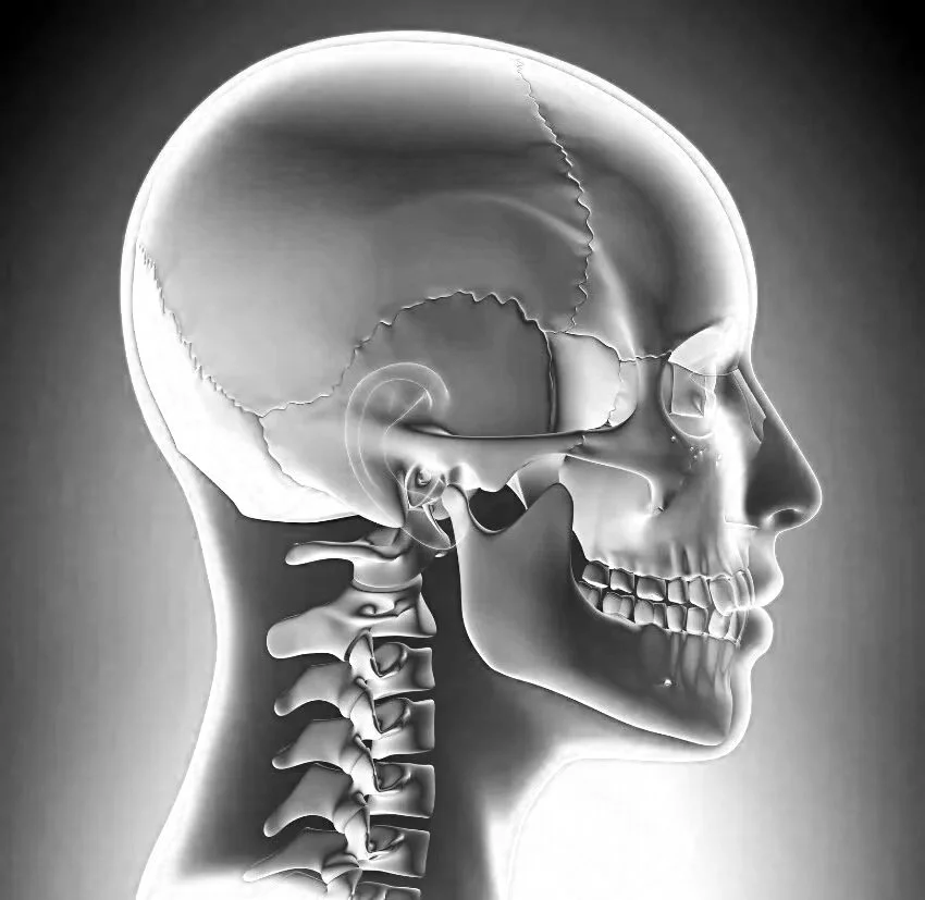
4. The Cranium
Wait — cranium… that’s the head, right? So how on earth could it possibly be connected to the sacrum?
I’m glad you asked, because this is where things become truly fascinating.
To understand this relationship, it helps to briefly step into the craniosacral perspective and the way it understands movement within the body. When I first learned anatomy, I was taught that the sacrum has three primary movements: flexion (nutation), extension (counter-nutation), and a subtle shifting forward and back.
Over time, my understanding has deepened. Through clinical practice and craniosacral work, the sacrum reveals itself as far more fluid, responsive, and expressive than those early lists suggest. For now, though, let’s stay with the classical movements of the sacrum:
The superior and anterior rotation (upward and forward) is the movement called nutation after the Latin word which means ‘to nod’. In this motion, the top of the sacrum comes into the true pelvis, i.e. birth canal. In some circles this movement is referred to as flexion of the sacrum.
The posterior and inferior rotation (backward and downward) movement is the opposite motion and it is referred to as counter-nutation or extension of the sacrum.
Slight movement along the sagittal plane.
The axis of the rotation is transverse at the level of the 2nd sacral segment. This is where it gets really interesting. The 2nd sacral segment happens to be the location where the spinal dura attaches. The spinal dura pulls the sacrum into flexion, and therefore creates a link between the sacrum and the occiput, where it also attaches. It is fascinating to visualise the movement of the sacrum influencing what happens in the cranium via this connection through the dura. The implication is that craniosacral and fascial therapy can positively influence the cranial bones through contact with the sacrum, and vice versa.
Sacral support, trauma, and regulation
In trauma-informed work, the sacrum often holds stories that were shaped long before words were available. When early experiences required the body to stay alert, braced, or prepared to adapt, the base of the spine can quietly carry that effort over time. Gentle support to the sacrum can invite the nervous system out of vigilance and into regulation, not by forcing release, but by offering a sense of safety and choice. As the sacrum softens, many people notice a deeper breath, steadier grounding, and a renewed capacity to feel supported from within. This is not about fixing the body, but about allowing it to remember how to rest.
Craniosacral therapy and scar work
In craniosacral therapy, the sacrum is approached with light, attentive touch, listening for subtle rhythms and patterns of tension within the body. This work supports the relationship between the sacrum, spine, and nervous system, often creating a sense of deep rest and integration.
Scar tissue, whether from surgery, injury, or birth, can sometimes disrupt how the sacrum and pelvis move and communicate with the rest of the body. Gentle scar work can help restore sensation, mobility, and coherence in these areas, supporting both physical comfort and nervous system regulation.
This kind of work is always guided by your body’s pace, capacity, and consent.
If you’re curious about how sacral support might feel in your own body, you’re warmly welcome to explore this work in a one-to-one session. Sessions are gentle, collaborative, and guided by your nervous system’s pace, with space to listen, settle, and restore a felt sense of support.
You don’t need to know what you’re coming for. We begin with what your body is already saying.
Available in Woodstock, Oxfordshire, and online for somatic support.
Sources:
Hugh Milne, The Heart of Listening. The Visionary Approach to Craniosacral Work, 1995
Frank Netter, Atlas of Human Anatomy, 2015
Andrew Biel, Trail Guide to the Body, 2013